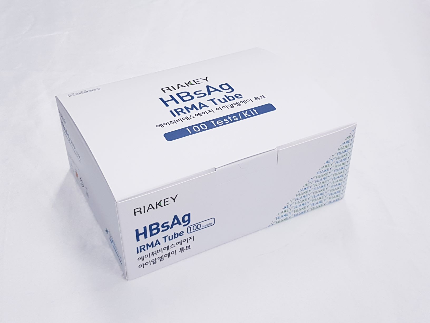
Product

RIAKEY HBsAg IRMA Tube
Intended Use
Immunoradiometric assay for quantitative determination of Hepatitis B surface Antigen (HBsAg) in human serum or plasma
Introduction
In 1965, Dr. Blumberg who was studying hemophilia, found an antibody in two patients which reacted against an antigen from an Australian Aborigine. Later the antigen was found in patients with serum type hepatitis and was initially designated "Australian Antigen". Subsequent study has shown the Australian Antigen to be the hepatitis B surface antigen (HBsAg). Initially there appeared to be three particles associated with hepatitis B infection: a large "complete" particle called the "Dane particle", an oblong 42nm particle and a small circular 22nm particle. Further research identified the Dane particle as the hepatitis B virion and the other two particles as excess surface protein. This former terminology is no longer used and the virus is referred to according to its structure. HBsAg presents an antigenic heterogeneity. The principle determinant is called “a” and is common to all the different types of HBsAg. There are two other pairs of major determinants that is d/y (1y, 2y, 3y) and w/r which are mutually exclusive. Therefore the following combinations are possible adw, adr, ayw, ayr. Subsequent association with hepatitis B virus (HBV) led to the development of sensitive, specific markers of HBV infection. During acute and chronic HBV infection, HBsAg is produced in excess amounts, circulating in blood as both 22nm spherical and tubular particles. HBsAg can be identified in serum 30~60 days after exposure to HBV and persists for variable periods depending on the resolution of the infection. Antibody to HBsAg (anti-HBs) develops after a resolved infection and is responsible for long-term immunity. Anti-HBc develops in both resolved acute infections and chronic HBV infections and persists indefinitely. Immunoglobulin M (IgM) anti-HBc appears early in infection and persists for greater than or equal to 6 month. It is a reliable marker of acute HBV infection.
Principle of the Assay
The RIAKEY HBsAg IRMA Tube is a non-competitive immunoradiometric (IRMA) method (“sandwich”). The method employs two highly specific monoclonal anti-HBs antibodies which recognize two different epitopes of the molecule. One antibody is coated on solid phase (coated tube), the other, specific for the HBsAg and labeled with Iodine-125, is used as a tracer. Antibody-coated polystyrene tubes serve as solid phase. The tracer antibody and the coated antibody react simultaneously with the HBsAg present in the standards, control serum and samples. Unbounded material is removed by a washing step. The amount of bound tracer will be directly proportional to the HBsAg concentration and the remaining radioactivity bound to the tubes is measured in a gamma scintillation counter.
- Do not use mixed reagents from different lots.
- Do not use reagents beyond the expiration date.
- Use distilled water stored in clean container.
- Use an individual disposable tip for each sample and reagent, to prevent the possible cross-contamination among the samples.
- Store the unused coated tubes at 2~8ºC in the appropriate bags with silica gel and accurately sealed.
- If large quantity of assay would be performed at one time, there might be substantial time variation between 60 tubes at one time to minimize time variation. Also, do not exceed 10 minutes for entire pipetting.
- Wear disposable globes while handling the kit reagents and wash hands thoroughly afterwards.
- Do not pipette by mouth.
- Do not smoke, eat or drink in areas where specimens or kit reagents are handle.
- Handle samples, reagents and loboratory equipments used for assy with extreme care, as they may potentially contain infectious agents.
- When samples or reagents happen to be split, wash carefully with a 3% sodium hypochlorite solution.
- Dispose of this cleaning liquid and also such used washing cloth or tissue paper with care, as they may also contain infectious agents.
- Avoid microbial contamination when the reagent vial be eventually opend or the contents be handled.
- Use only for IN VITRO.

