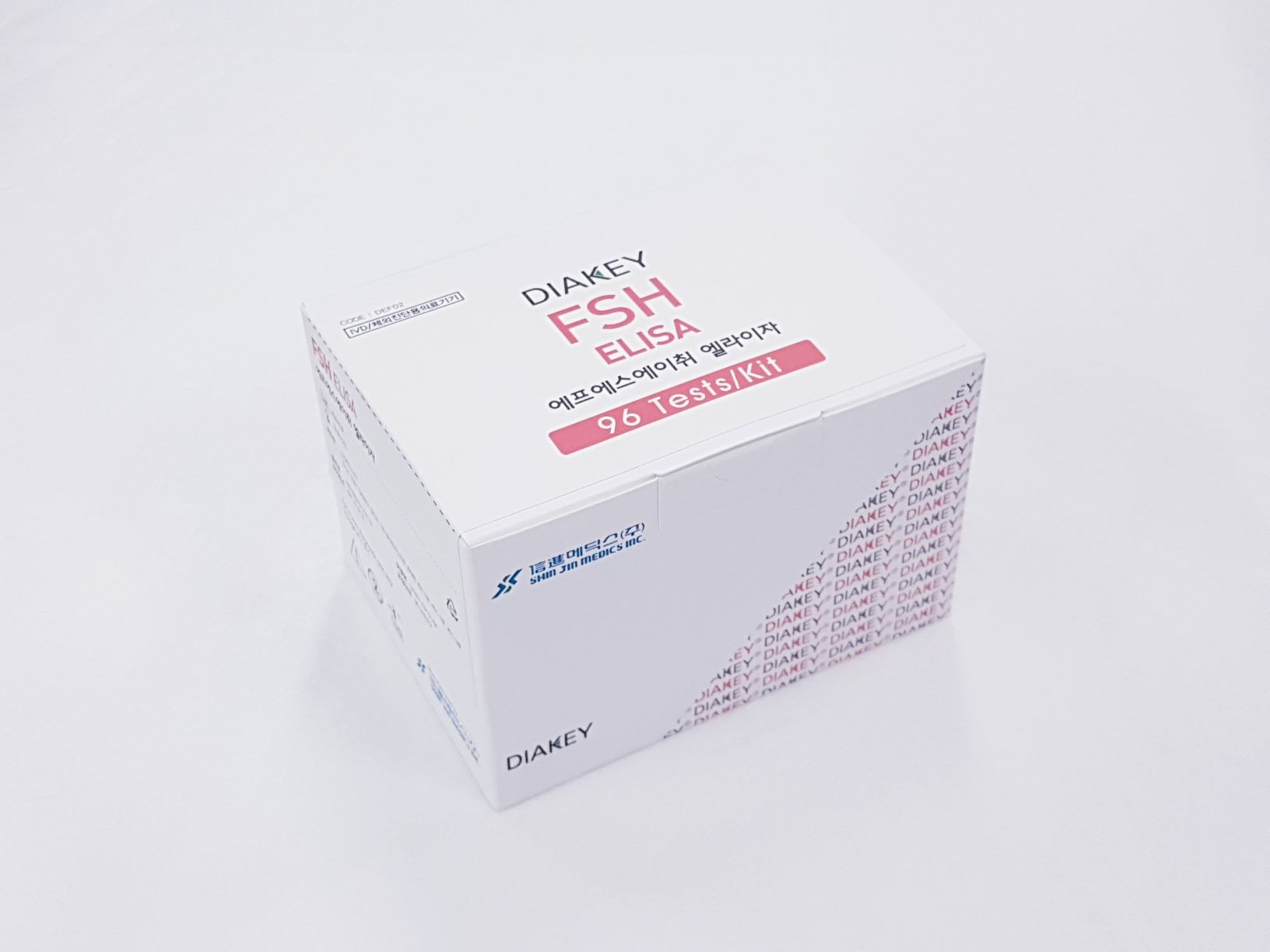
Product

DIAKEY FSH ELISA
Intended Use
Enzyme immunoassay for quantitative determination of follicle-stimulating hormone (FSH) in human serum or plasma
Introduction
Follicle stimulating hormone (FSH) is secreted by the β-cells of the anterior pituitary under the control of the gonadotropin releasing hormone produced in the hypothalamus. It is a glycoprotein with a molecular weight of approximately 28,000 consisting of two different polypeptide subunits designated alpha (α) and beta (β) which are associated by not covalent binding. The subunit alpha (α) compound by 89 amino acids, has a similar structure as other glycoprotein hormone (FSH, LH, TSH and HCG). The subunit beta (β), a specific hormone, gives the immunological feature to the whole molecule. The alpha (α) chains of FSH, LH, TSH and HCG are biochemically identical, whereas the beta (β) chains are biochemically unique and confer biological and immunological specificity. Bioactivity is also determined by the beta (β) chain. The FSH is produced by the basophil cells of the frontal lobe of hypophysis. The secretion is stimulated by the hypophysis glands are the gonadotropin releasing hormone (GnRH). The FSH facilitates the development and maintenance of gonadal tissues, which synthesize and secrete steroid hormones. The activity of the hypothalamic and hypophysis glands are controlled by the concentration of the circulating steroids performs the decrease or increase of FSH (negative feedback). Circulating level of FSH is controlled by a negative feedback effect on the hypothalamus by steroidal hormones.
Principle of the Assay
The DIAKEY FSH ELISA is a solid-phase, non-competitive immunoassay based upon the sandwich technique. Two different monoclonal anti-FSH antibodies are used. One antibody is coated on solid phase (coated plate), the other, specific for the FSH and labeled with HRP is used as a conjugate. The conjugate antibody and the coated antibody react simultaneously with the FSH antigen present in the standards, control serum and sample. Unbounded material is removed by a washing step. After washing, substrate reagent is added to each well and the enzyme reaction is allowed to proceed. During the enzyme reaction a blue color will develop if antigen is present. The intensity of the color is proportional to the amount of FSH present in the samples. The color intensity is determined in a microplate spectrophotometer at 450nm (reference 620nm).
Handling Precaution
- Do not use mixed reagents from different lots.
- Do not use reagents beyond the expiration date.
- Use distilled water stored in clean container.
- Use an individual disposable tip for each sample and reagent, to prevent the possible cross-contamination among the samples.
- Rapidly dispense reagents during the assay, not to let wells dry out.
Use Precaution
- Wear disposable globes while handling the kit reagents and wash hands thoroughly afterwards.
- Do not pipette by mouth.
- Do not smoke, eat or drink in areas where specimens or kit reagents are handle.
- Handle samples, reagents and loboratory equipments used for assy with extreme care, as they may potentially contain infectious agents.
- When samples or reagents happen to be spilt, wash carefully with a 1% sodium hypochlorite solution.
- Dispose of this cleaning liquid and also such used washing cloth or tissue paper with care, as they may also contain infectious agents.
- Avoid microbial contamination when the reagent vial be eventually opend or the contents be handled.
- Use only for IN VITRO.

