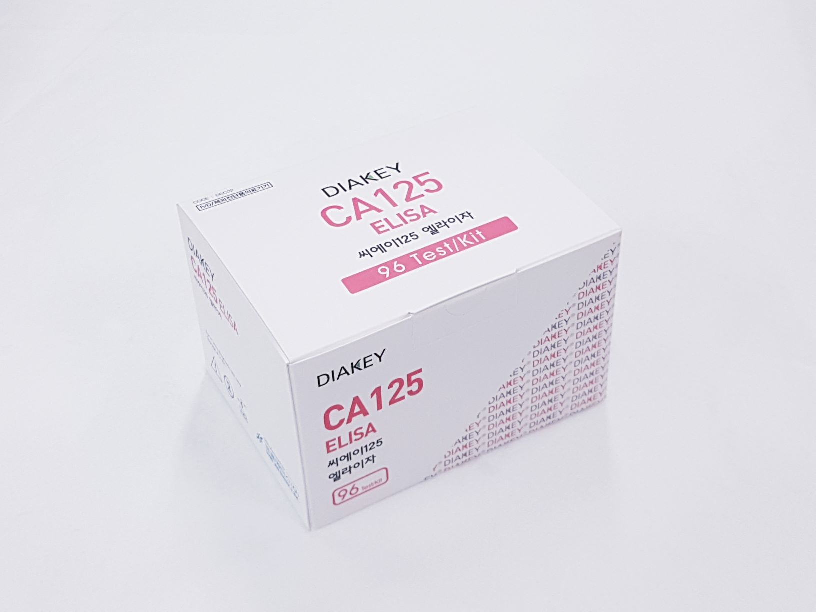
Product

DIAKEY CA125 ELISA
Intended Use
Enzyme immunometric assay for quantitative determination of cancer antigen125 (CA125) in human serum or plasma
Introduction
In 1981, Bast et al, first obtained monoclonal antibody CA125, which was produced using lymphocytes from a mouse immunized with OVCA 433, a cell line derived from a papillary serous cystadenocarcinoma of the ovary. The CA125 reactive determinants, isolated from either cell culture or serum, are found on a heterogeneous, high molecular weight (200 to 1,000 kilo Dalton) glycoprotein (designated CA125 antigen). The determinant is proteinaceous in nature, but has nondeterminant associated carbohydrate moieties. CA125 reactive determinants are not present in the sur face epithelium of either normal fetal or adult ovaries, with the exception of inclusion cysts, areas of metaplasia, and papillary excrescences. In fetal tissue, CA125 reactive determinants have been observed in the amnion and in derivatives of the coelomic epithelium. In women with primary epithelial ovarian carcinoma who had undergone first-line therapy and were candidates for diagnostic second-look procedures, a CA125 assay value greater than or equal to 35 U/㎖ was found to be indicative of the presence of residual tumor. Assuming the physician cannot identify alternative causes for an elevated CA125 assay value, a CA125 assay value determined to be greater than or equal to 35 U/㎖ provides substantial evidence that residual tumor is present.
Principle of the Assay
The DIAKEY CA125 ELISA is a solid-phase, non-competitive immunoassay based upon the sandwich technique. Two different monoclonal anti-CA125 antibodies are used. One antibody is coated on solid phase (coated plate), the other, specific for the CA125 and labeled with HRPO, is used as a conjugate. The conjugate antibody and the coated antibody react simultaneously with the CA125 antigen present in the standards, control serum and samples. Unbounded material is removed by a washing step. After washing, substrate reagent is added to each well and the enzyme reaction is allowed to proceed. During the enzyme reaction a blue color will develop if antigen is present. The intensity of the color is proportional to the amount of CA125 present in the samples. The color intensity is determined in a microplate spectrophotometer at 450nm (reference 620nm).
Handling Precaution
- Do not use mixed reagents from different lots.
- Do not use reagents beyond the expiration date.
- Use distilled water stored in clean container.
- Use an individual disposable tip for each sample and reagent, to prevent the possible cross-contamination among the samples.
- Rapidly dispense reagents during the assay, not to let wells dry out.
Use Precaution
- Wear disposable globes while handling the kit reagents and wash hands thoroughly afterwards.
- Do not pipette by mouth.
- Do not smoke, eat or drink in areas where specimens or kit reagents are handle.
- Handle samples, reagents and loboratory equipments used for assy with extreme care, as they may potentially contain infectious agents.
- When samples or reagents happen to be spilt, wash carefully with a 1% sodium hypochlorite solution.
- Dispose of this cleaning liquid and also such used washing cloth or tissue paper with care, as they may also contain infectious agents.
- Avoid microbial contamination when the reagent vial be eventually opend or the contents be handled.
- Use only for IN VITRO.

