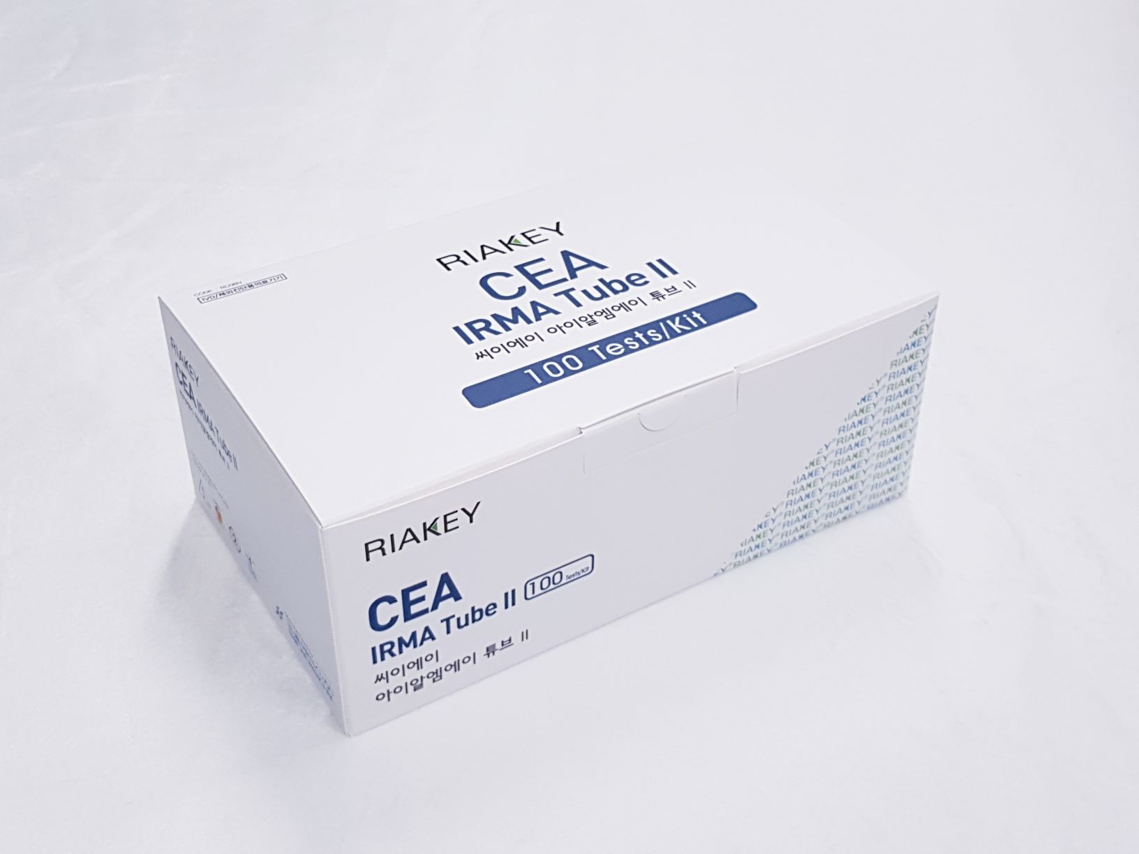
Product

RIAKEY CEA IRMA Tube II
Intended Use
Immunoradiometric assay for quantitative determination of carcinoembryonic antigen (CEA) in human serum or plasma
Introduction
The carcinoembryonic antigen (CEA) is a glycoprotein soluble in perchloric acid, first identified by Gold and Freedman in embryonic endodermal tissues of 2-6 month old feti and in epithelial tumors of endodermal origin (gastrointestinal tract). CEA molecule presents a high degree of heterogeneity, depending on the carbohydrate contents (50-60%) and also influenced by the purification method. Heterogeneity results clear also from molecular weight (175,000-2,000,000 daltons), sedimentation coefficient (6.2-6.8 S) and isoelectric point (p.l. 3-4), while it’s electrophoretic mobility is always beta. CEA immunological determination allowed evidencing of CEA-like molecules, a family of molecules with common antigenic determinants, synthesized by normal and pathological tissues, among which the most relevant one is NCA (Non-specific cross reacting antigen). Use of monoclonal antibodies has made it possible to definitely overcome the problem of the cross-reacting CEA like molecules when assaying CEA.
Principle of the Assay
The RIAKEY CEA IRMA Tube II is an one step non-competitive immunoradiometric (IRMA) method (“sandwich”). The method employs two highly specific monoclonal anti-CEA antibodies which recognize two different epitopes of the molecule. One antibody is coated on solid phase (coated tube), the other, specific for the CEA and labeled with Iodine-125, is used as a tracer. Antibody-coated polystyrene tubes serve as solid phase. The tracer antibody and the coated antibody react simultaneously with the CEA antigen present in the standards, control serum and samples. Unbounded material is removed by a washing step. The amount of bound tracer will be directly proportional to the CEA antigen concentration and the remaining radioactivity bound to the tubes is measured in a gamma scintillation counter.
- Do not use mixed reagents from different lots.
- Do not use reagents beyond the expiration date.
- Use distilled water stored in clean container.
- Use an individual disposable tip for each sample and reagent, to prevent the possible cross-contamination among the samples.
- Store the unused coated tubes at 2~8ºC in the appropriate bags with silica gel and accurately sealed.
- If large quantity of assay would be performed at one time, there might be substantial time variation between 60 tubes at one time to minimize time variation. Also, do not exceed 10 minutes for entire pipetting.
- Wear disposable globes while handling the kit reagents and wash hands thoroughly afterwards.
- Do not pipette by mouth.
- Do not smoke, eat or drink in areas where specimens or kit reagents are handle.
- Handle samples, reagents and loboratory equipments used for assy with extreme care, as they may potentially contain infectious agents.
- When samples or reagents happen to be split, wash carefully with a 3% sodium hypochlorite solution.
- Dispose of this cleaning liquid and also such used washing cloth or tissue paper with care, as they may also contain infectious agents.
- Avoid microbial contamination when the reagent vial be eventually opend or the contents be handled.
- Use only for IN VITRO.

