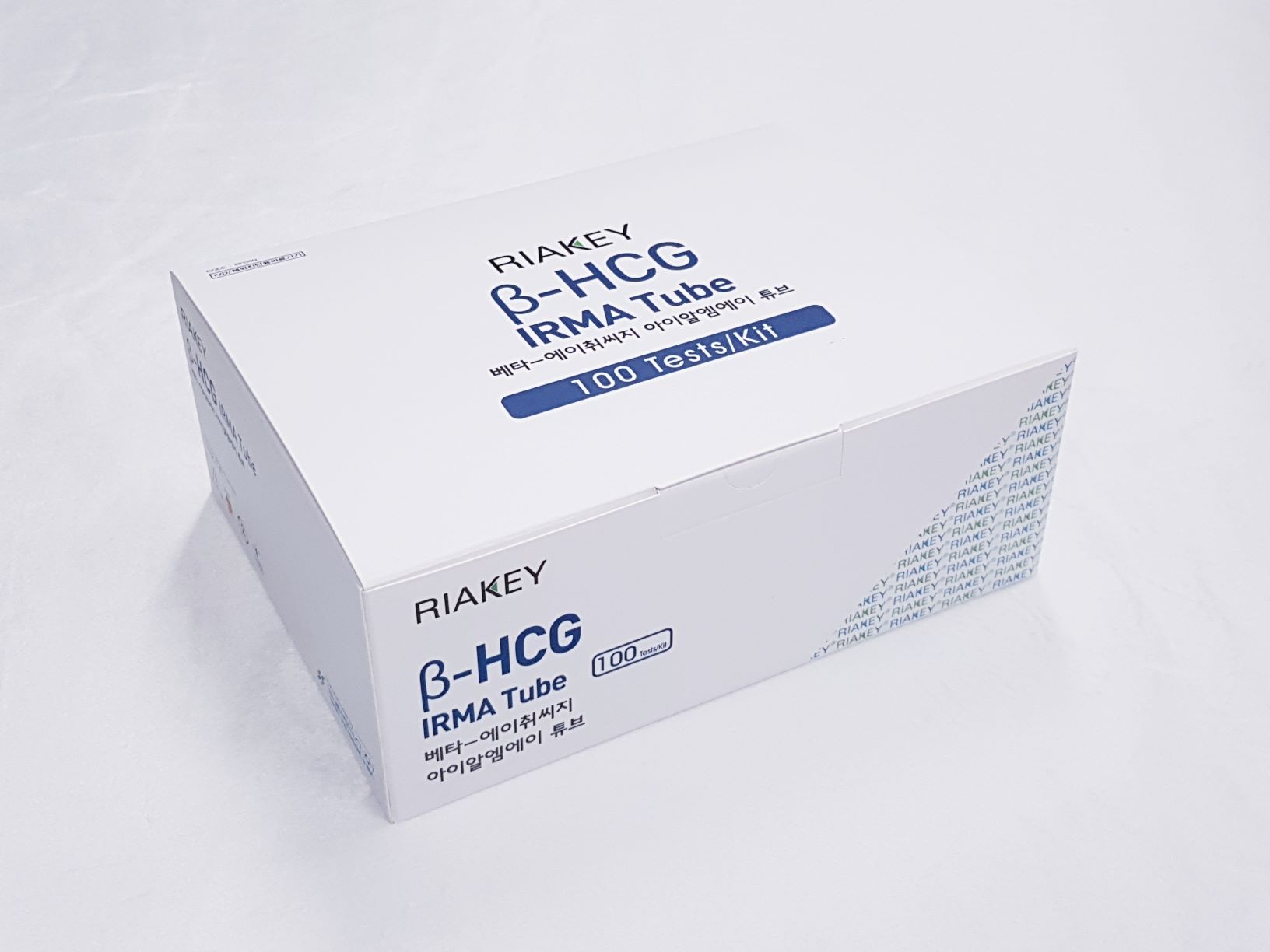
Product

RIAKEY β-hCG IRMA Tube
Intended Use
Immunoradiometric assay for quantitative determination of Chorionic Gonadotropin (hCG) in human serum or plasma
Introduction
Human Chorionic Gonadotropin (hCG) is a glycoprotein (molecular weight: 38000 Dalton) with approximately 30% carbohydrate content and 9% of sialic acid; it is secreted during pregnancy, by syncytiotrophoblast cells of the placenta. The molecule is composed of two subunits known as αand β. The α-subunit shares structural homology with pituitary glycoprotein hormones (FSH, LH and TSH). The β-subunit is characterized by a sequence of 30 amino acids at the C-terminal, which are specific for each hormone. In this way, unique biological as well as immunological activity is conferred to each hormone. Thus, a specific hCG assay is only possible by means of antibodies directed against the β-subunit. The most important function of hCG is to sustain the corpus luteum in producing Estradiol and Progesterone, thereby supporting pregnancy, in the male fetus in addition, it stimulates Leydig cells to produce Testosterone, thus contributing to sexual differentiation.
Principle of the Assay
The present method is based upon two anti-βhCG monoclonal antibodies recognizing two different epitopes of the molecule. One antibody is adsorbed to the solid phase (coated tube), the other-labeled with Iodine-125 is used as tracer, and the sample to be tested is first incubated in the coated tube. Following this incubation and after an aspirating / washing cycle, the labeled antibody is added to each coated tube, where it is bound to the solid phase by means of the antigen in the standards and (if present) in samples. The amount of binding will be directly proportional to the antigen concentration. After tracer incubation, another washing / aspirating cycle will remove any liquid reagent residue. The radioactivity in the tubes is measured in a gamma counter.
- Do not use mixed reagents from different lots.
- Do not use reagents beyond the expiration date.
- Use distilled water stored in clean container.
- Use an individual disposable tip for each sample and reagent, to prevent the possible cross-contamination among the samples.
- Store the unused coated tubes at 2~8ºC in the appropriate bags with silica gel and accurately sealed.
- If large quantity of assay would be performed at one time, there might be substantial time variation between 60 tubes at one time to minimize time variation. Also, do not exceed 10 minutes for entire pipetting.
- Wear disposable globes while handling the kit reagents and wash hands thoroughly afterwards.
- Do not pipette by mouth.
- Do not smoke, eat or drink in areas where specimens or kit reagents are handle.
- Handle samples, reagents and loboratory equipments used for assy with extreme care, as they may potentially contain infectious agents.
- When samples or reagents happen to be split, wash carefully with a 3% sodium hypochlorite solution.
- Dispose of this cleaning liquid and also such used washing cloth or tissue paper with care, as they may also contain infectious agents.
- Avoid microbial contamination when the reagent vial be eventually opend or the contents be handled.
- Use only for IN VITRO.

