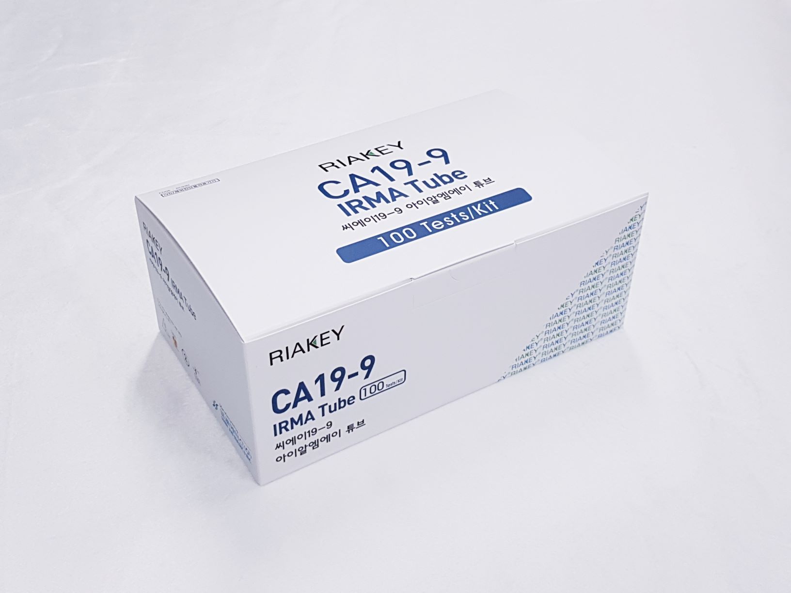
Product

RIAKEY CA19-9 IRMA Tube II
Intended Use
Immunoradiometric assay for quantitative determination of carbohydrate antigen 19-9(CA19-9) in human serum or plasma
Introduction
Macromolecular tumor markers can be classified into the following major classes: enzymes, hormones and growth factors and their receptors, oncogenic products, viral antigens, oncofetal and surface antigens. With a few exceptions (certain enzymes), most macromolecular tumor markers are glycol-conjugates and a wealth of information is available on these antigens. Therefore, investigations into the expressions of unusual glycoprotein antigens ('markers') associated with tumors and their metastases dominate much of clinical and experimental oncology today. CA19-9 antigen is a kind of carbohydrate antigen, one of important tumor markers. The monoclonal antibody was obtained by Koprowski et al, in 1979 from hybrids of human colon cancer cell line cultured in vitro. The determinant of CA19-9 was proved to be a sialylated lacto-N-fucopentaose Ⅱ. Its biochemical properties are associated with lewis blood group antigens. Analyses of serum samples by several groups have indicated elevated levels of CA19-9 antigen in several types of malignancies, particularly those of pancreas, stomach and colon, but not in sera of normal individuals. CA19-9 antigen was originally identified as a ganglioside isolated from the SW1116 human colorectal carcinoma cell line, while the serum antigen was characterized as a mucin. It can also be detected on mucin in patients' sera. Sources include normal pancreas, bile duct, and gastric, colic, endometric and salivary epithelia. In healthy individuals, the CA19-9 antigen circulates at low levels, normally less than 37U/mL. Elevated levels have been seen in benign inflammatory diseases of the hepatobiliary tract. Studies have also shown that mucin bearing this antigen was more frequently detected in the sera of patients with pancreatic cancer than any other gastrointestinal carcinoma, including colorectal. It is also found in bile duct, ovarian mucinous cystadenocarcinomas and uterine adenocarcinomas.
Principle of the Assay
The assay is a non-competitive immunoradiometric assay (IRMA) method (sandwich principle). The present method employs two monoclonal anti-CA19-9 antibodies which recognize two different epitopes of the molecules. One antibody is absorbed in solid phase (coated tube), the other (labeled with Iodine-125) is used as tracer. The sample to be tested, is incubated in the coated tube, following the incubation, after aspiration and washing, the labeled antibody is added to the coated tubes, where it binds to the solid phase, by means of the antigen in standards and samples. The amount of bound tracer will thus be directly proportional to the antigen concentration. After a further aspiration and washing cycle, the residual radioactivity in the tubes is measured in a gamma counter.
- Do not use mixed reagents from different lots.
- Do not use reagents beyond the expiration date.
- Use distilled water stored in clean container.
- Use an individual disposable tip for each sample and reagent, to prevent the possible cross-contamination among the samples.
- Store the unused coated tubes at 2~8ºC in the appropriate bags with silica gel and accurately sealed.
- If large quantity of assay would be performed at one time, there might be substantial time variation between 60 tubes at one time to minimize time variation. Also, do not exceed 10 minutes for entire pipetting.
- Wear disposable globes while handling the kit reagents and wash hands thoroughly afterwards.
- Do not pipette by mouth.
- Do not smoke, eat or drink in areas where specimens or kit reagents are handle.
- Handle samples, reagents and loboratory equipments used for assy with extreme care, as they may potentially contain infectious agents.
- When samples or reagents happen to be split, wash carefully with a 3% sodium hypochlorite solution.
- Dispose of this cleaning liquid and also such used washing cloth or tissue paper with care, as they may also contain infectious agents.
- Avoid microbial contamination when the reagent vial be eventually opend or the contents be handled.
- Use only for IN VITRO.

